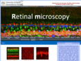
Retinal microscopy The retina is a translucent filmy piece of tissue lining the back of the eyeball. The multiple layers of interconnected neurons in this thin slab of neural tissue are in charge of the first of steps of vision. Cones and rods absorb the incident light. The phototransduction mechanisms housed in the outer segments of these cells, transduce light into electrical signals that are relayed on to bipolar cells, in the first synaptic layer of the retina called outer plexiform layer. Visual information is processed further at the following layer where amacrine cells form a second complex network of synaptic interconnections the inner plexiform layer. Lastly, visual information is conveyed to the ganglion cells and exits the eye along the optic nerve in its way to the brain. Each species have the retina adapted to light conditions dependent on whether the animal lives in diurnal or nocturnal habitats. Retinalmicroscopy.com~Site InfoWhoisTrace RouteRBL Check
Virtual Microscopy Bamras-VM is an internet-based virtual microscopy of opportunistic microorganisms from clinical specimens (opportunistic infection). The image can be moved (left-right, up and down) and magnified like examining under a microscope. Bamras-eqa.com~Site InfoWhoisTrace RouteRBL Check